Home

Formazione conchiglia primavera pkh26 staining Consigliere rivista Senatore

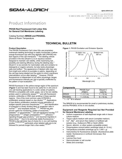
PKH26 Red Fluorescent Cell Membrane Labeling Sigma-Aldrich

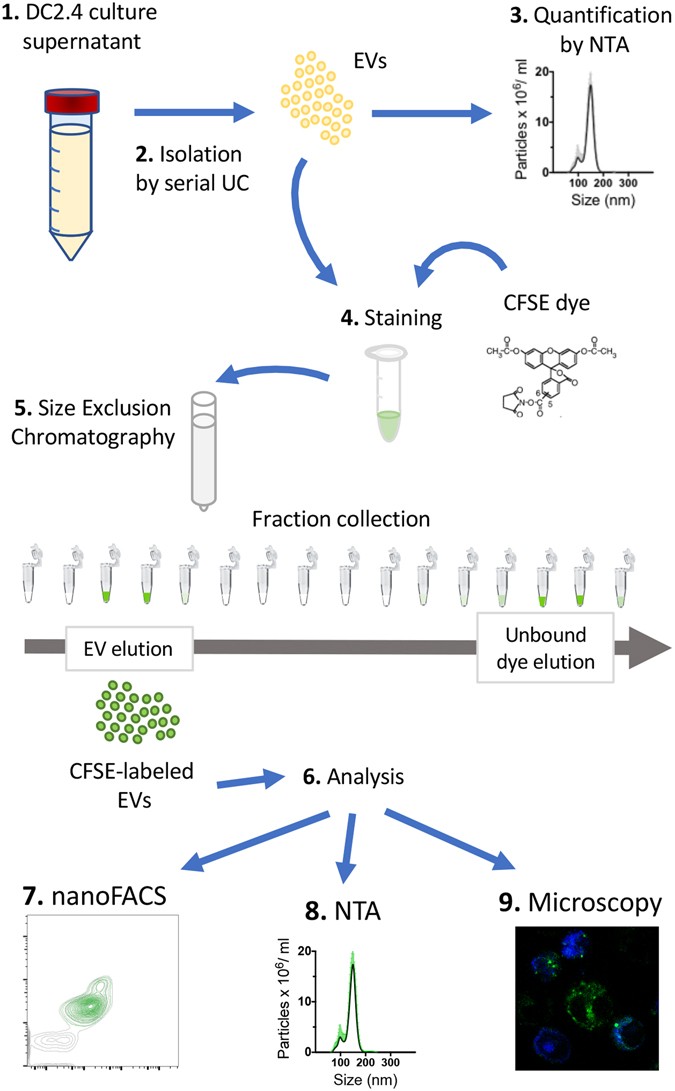
Labeling Extracellular Vesicles for Nanoscale Flow Cytometry | Scientific  Reports
Labeling Extracellular Vesicles for Nanoscale Flow Cytometry | Scientific Reports

Standard protocol for PKH26 dye labeling (Sigma, Aldrich) | Download  Scientific Diagram
Standard protocol for PKH26 dye labeling (Sigma, Aldrich) | Download Scientific Diagram

a ) Representative scan of PKH26-labelled bovine mesenchymal stem... |  Download Scientific Diagram
a ) Representative scan of PKH26-labelled bovine mesenchymal stem... | Download Scientific Diagram

PKH26 Can Transfer to Host Cells In Vitro and Vivo | Stem Cells and  Development
PKH26 Can Transfer to Host Cells In Vitro and Vivo | Stem Cells and Development

Extracellular vesicles from human stem cells can alleviate cellular aging  in culture | Exosome RNA
Extracellular vesicles from human stem cells can alleviate cellular aging in culture | Exosome RNA

Detection of plasma membrane fragments exchange by PKH26 and PKH67 dyes.

Optimized staining and proliferation modeling methods for cell division  monitoring using cell tracking dyes. - Abstract - Europe PMC
Optimized staining and proliferation modeling methods for cell division monitoring using cell tracking dyes. - Abstract - Europe PMC

New Lipophilic Fluorescent Dyes for Exosome Labeling: Monitoring of  Cellular Uptake of Exosomes | bioRxiv
New Lipophilic Fluorescent Dyes for Exosome Labeling: Monitoring of Cellular Uptake of Exosomes | bioRxiv

LumiTrace PKH26 cell membrane labeling kit

PKH26 Red Fluorescent Cell Membrane Labeling Sigma-Aldrich

BDNF-Hypersecreting Human Mesenchymal Stem Cells Promote Functional  Recovery, Axonal Sprouting, and Protection of Corticospinal Neurons after  Spinal Cord Injury | Journal of Neuroscience
BDNF-Hypersecreting Human Mesenchymal Stem Cells Promote Functional Recovery, Axonal Sprouting, and Protection of Corticospinal Neurons after Spinal Cord Injury | Journal of Neuroscience
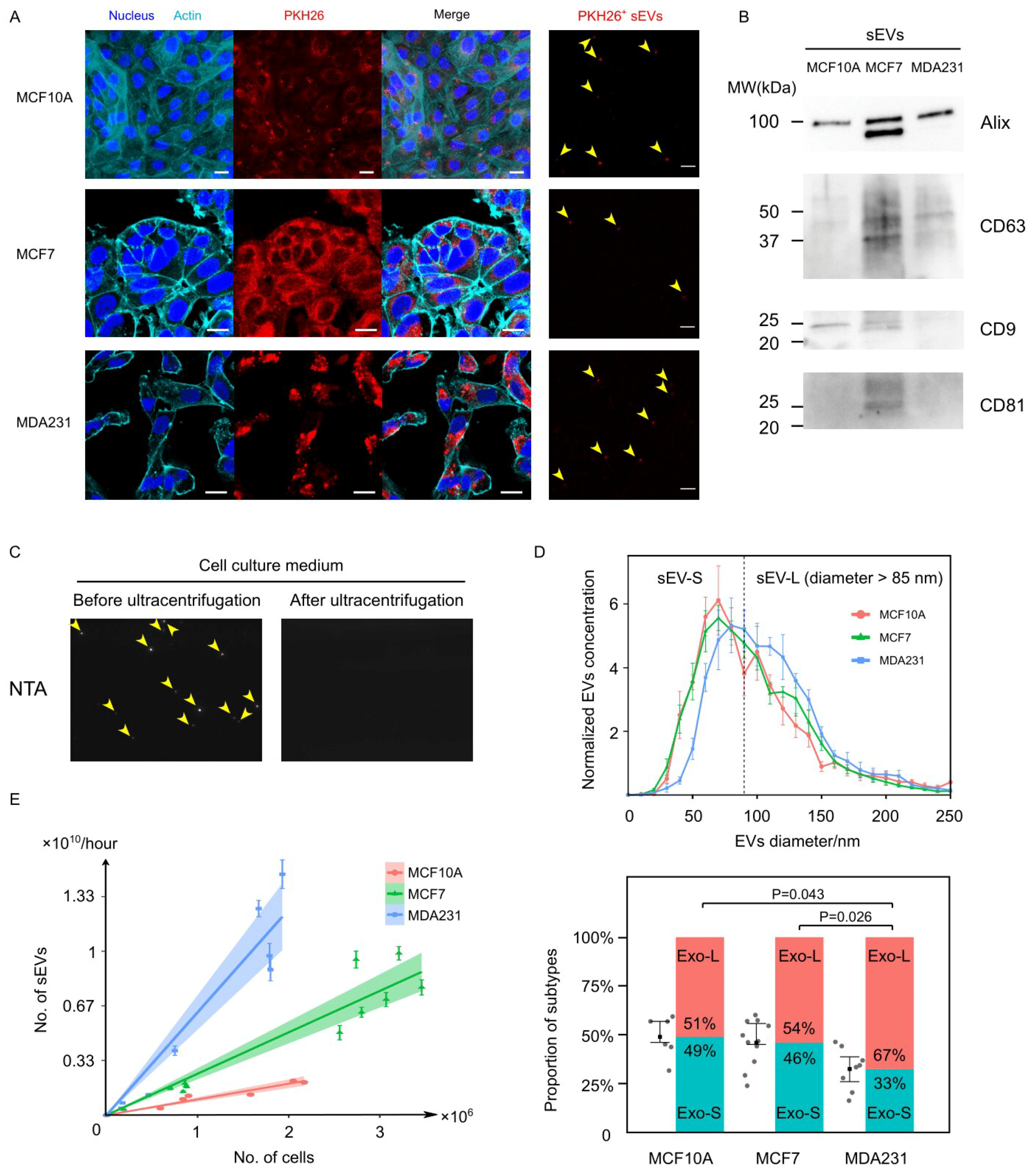
Cancers | Free Full-Text | Distinct mRNAs in Cancer Extracellular Vesicles  Activate Angiogenesis and Alter Transcriptome of Vascular Endothelial Cells
Cancers | Free Full-Text | Distinct mRNAs in Cancer Extracellular Vesicles Activate Angiogenesis and Alter Transcriptome of Vascular Endothelial Cells
Confocal optical sections of HeLa cells; the labelling with red... |  Download Scientific Diagram
Confocal optical sections of HeLa cells; the labelling with red... | Download Scientific Diagram

Fields of Interest - PKH2

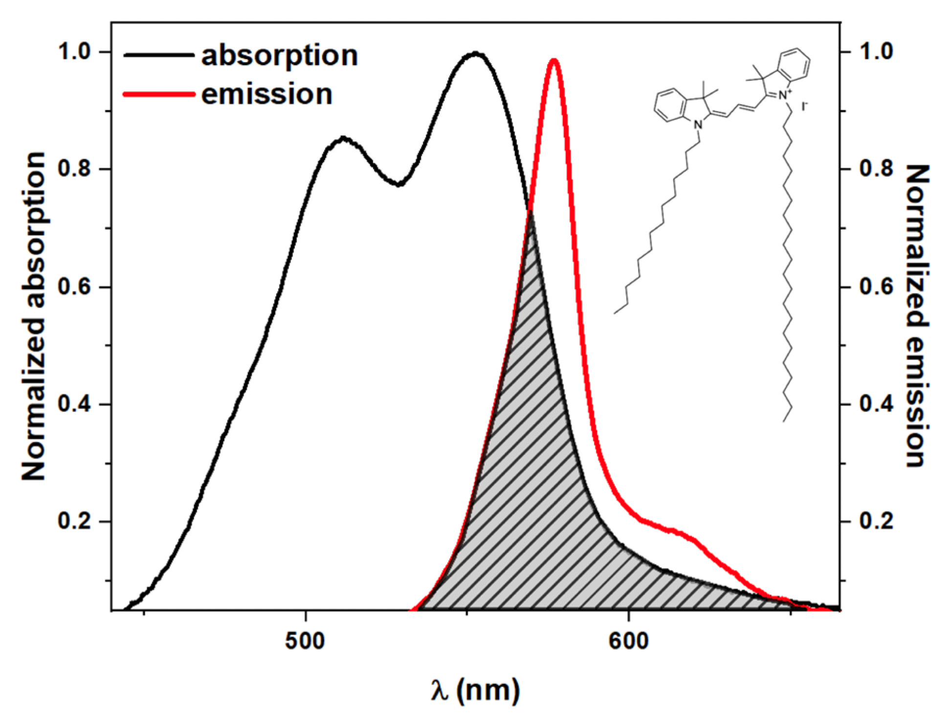
IJMS | Free Full-Text | Probing Red Blood Cell Membrane Microviscosity  Using Fluorescence Anisotropy Decay Curves of the Lipophilic Dye PKH26
IJMS | Free Full-Text | Probing Red Blood Cell Membrane Microviscosity Using Fluorescence Anisotropy Decay Curves of the Lipophilic Dye PKH26

Time‐lapse microscopy of macrophages during embryonic vascular development  - Al‐Roubaie - 2012 - Developmental Dynamics - Wiley Online Library
Time‐lapse microscopy of macrophages during embryonic vascular development - Al‐Roubaie - 2012 - Developmental Dynamics - Wiley Online Library

Optimized Staining and Proliferation Modeling Methods for Cell Division  Monitoring using Cell Tracking Dyes | Protocol
Optimized Staining and Proliferation Modeling Methods for Cell Division Monitoring using Cell Tracking Dyes | Protocol

Photomicrographs of PKH26 fluorescent-stained sections showing a... |  Download Scientific Diagram
Photomicrographs of PKH26 fluorescent-stained sections showing a... | Download Scientific Diagram

Design and Applications of a Fluorescent Labeling Technique for Lipid and  Surfactant Preformed Vesicles
Design and Applications of a Fluorescent Labeling Technique for Lipid and Surfactant Preformed Vesicles
A) The representative of PKH-26 label (B) The staining for nuclei... |  Download Scientific Diagram
A) The representative of PKH-26 label (B) The staining for nuclei... | Download Scientific Diagram

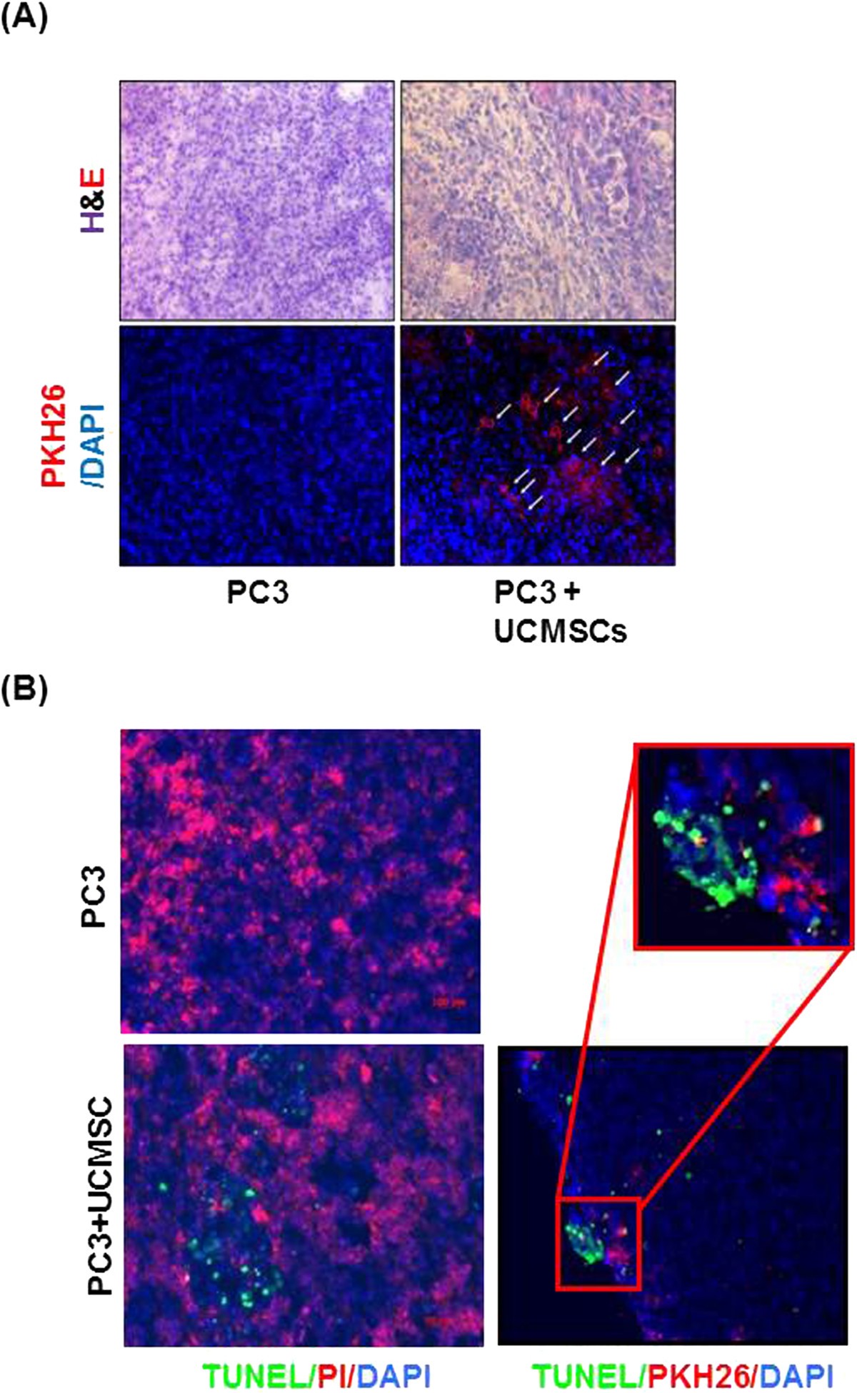
RETRACTED ARTICLE: Umbilical cord tissue-derived mesenchymal stem cells  induce apoptosis in PC-3 prostate cancer cells through activation of JNK  and downregulation of PI3K/AKT signaling | Stem Cell Research & Therapy |  Full
RETRACTED ARTICLE: Umbilical cord tissue-derived mesenchymal stem cells induce apoptosis in PC-3 prostate cancer cells through activation of JNK and downregulation of PI3K/AKT signaling | Stem Cell Research & Therapy | Full

PKH26 Staining Defines Distinct Subsets of Normal Human Colon Epithelial  Cells at Different Maturation Stages | PLOS ONE
PKH26 Staining Defines Distinct Subsets of Normal Human Colon Epithelial Cells at Different Maturation Stages | PLOS ONE

PKH26 Red Fluorescent Cell Linker Kit (MINI26 ... - Sigma-Aldrich

PKH26 Red Fluorescent Cell Lin | MINI26-1KT | SIGMA ALDRICH | SLS Ireland