Home

Senza fluente Piedi morbidi nissl staining neurons mar Mediterraneo Bambini scudo

Nissl Staining - an overview | ScienceDirect Topics

xmlinkhub
xmlinkhub

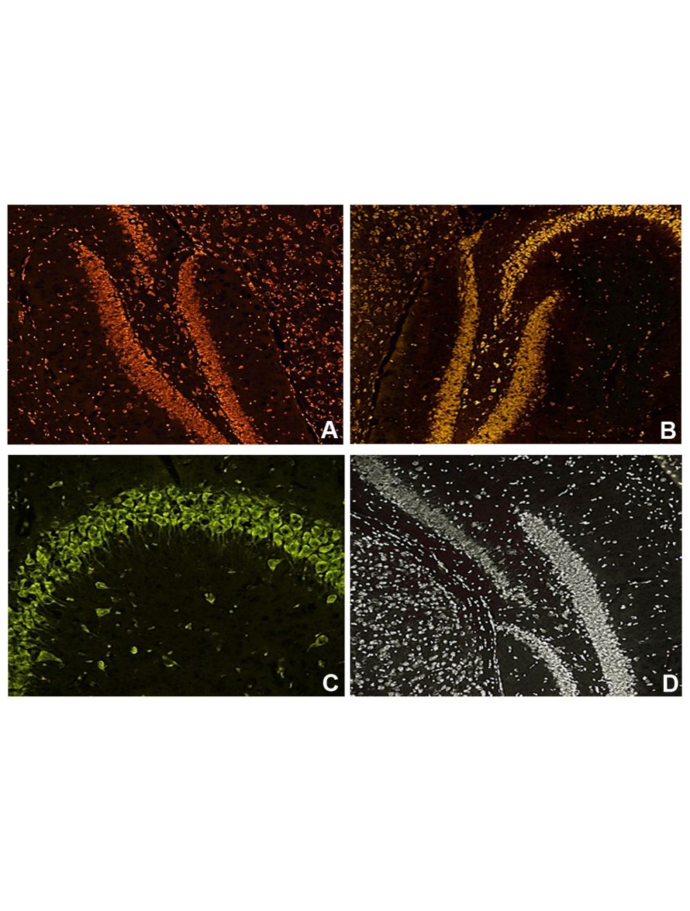
NisslFluor™ Nissl Sample Pack, Ready-to-Dilute™, Fluorescent Nissl Stains  for Healthy Neurons
NisslFluor™ Nissl Sample Pack, Ready-to-Dilute™, Fluorescent Nissl Stains for Healthy Neurons

Nissl Body - an overview | ScienceDirect Topics

Nissl staining of rat brain sections at different time‑points (x400... |  Download Scientific Diagram
Nissl staining of rat brain sections at different time‑points (x400... | Download Scientific Diagram

SciELO - Brasil - Franz Nissl (1860-1919), noted neuropsychiatrist and  neuropathologist, staining the neuron, but not limiting it Franz Nissl  (1860-1919), noted neuropsychiatrist and neuropathologist, staining the  neuron, but not limiting it
SciELO - Brasil - Franz Nissl (1860-1919), noted neuropsychiatrist and neuropathologist, staining the neuron, but not limiting it Franz Nissl (1860-1919), noted neuropsychiatrist and neuropathologist, staining the neuron, but not limiting it

Ischemia-induced neuronal cell death is mediated by the endoplasmic  reticulum stress pathway involving CHOP | Cell Death & Differentiation
Ischemia-induced neuronal cell death is mediated by the endoplasmic reticulum stress pathway involving CHOP | Cell Death & Differentiation

NeuroAnatomy 5 Flashcards | Quizlet

PDF] Constructing software for analysis of neuron, glial and endothelial  cell numbers and density in histological Nissl-stained rodent brain tissue  | Semantic Scholar
PDF] Constructing software for analysis of neuron, glial and endothelial cell numbers and density in histological Nissl-stained rodent brain tissue | Semantic Scholar

What is a Nissl stain? — Brain Stuff

Nissl stain analysis. A: After Nissl staining, the typical morphology... |  Download Scientific Diagram
Nissl stain analysis. A: After Nissl staining, the typical morphology... | Download Scientific Diagram
J Neurol Res
J Neurol Res

NisslFluor™ Red, Ready-to-Dilute™, Fluorescent Nissl Stain for Healthy  Neurons
NisslFluor™ Red, Ready-to-Dilute™, Fluorescent Nissl Stain for Healthy Neurons

Nissl staining of neuronal cells in the brain of neonatal offspring rats  from different groups.
Nissl staining of neuronal cells in the brain of neonatal offspring rats from different groups.

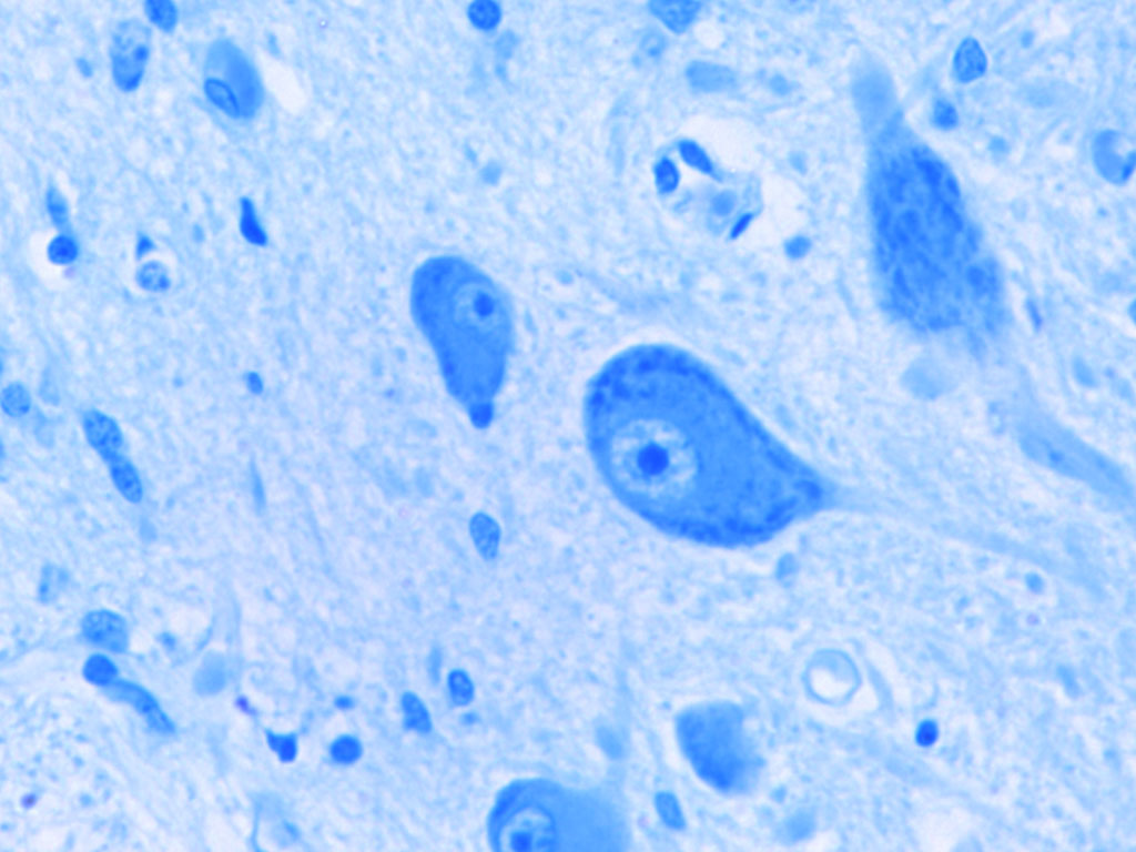
Motor Neurons. Nissl Bodies Stock Photo - Image of neurohistology, cord:  117240486
Motor Neurons. Nissl Bodies Stock Photo - Image of neurohistology, cord: 117240486

Nuclear chromatin patterns shown by Nissl staining are related to... |  Download Scientific Diagram
Nuclear chromatin patterns shown by Nissl staining are related to... | Download Scientific Diagram

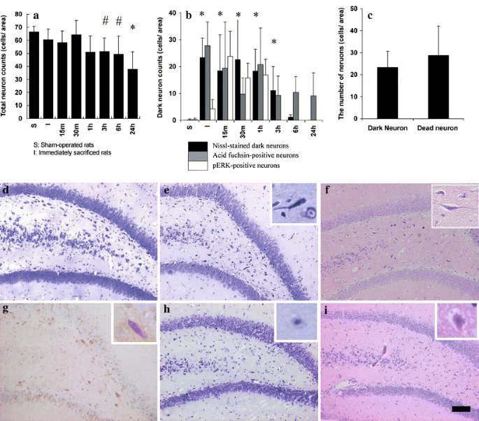
The fate of Nissl-stained dark neurons following traumatic brain injury in  rats: difference between neocortex and hippocampus regarding survival rate  | Acta Neuropathologica
The fate of Nissl-stained dark neurons following traumatic brain injury in rats: difference between neocortex and hippocampus regarding survival rate | Acta Neuropathologica

Histological methods for CNS

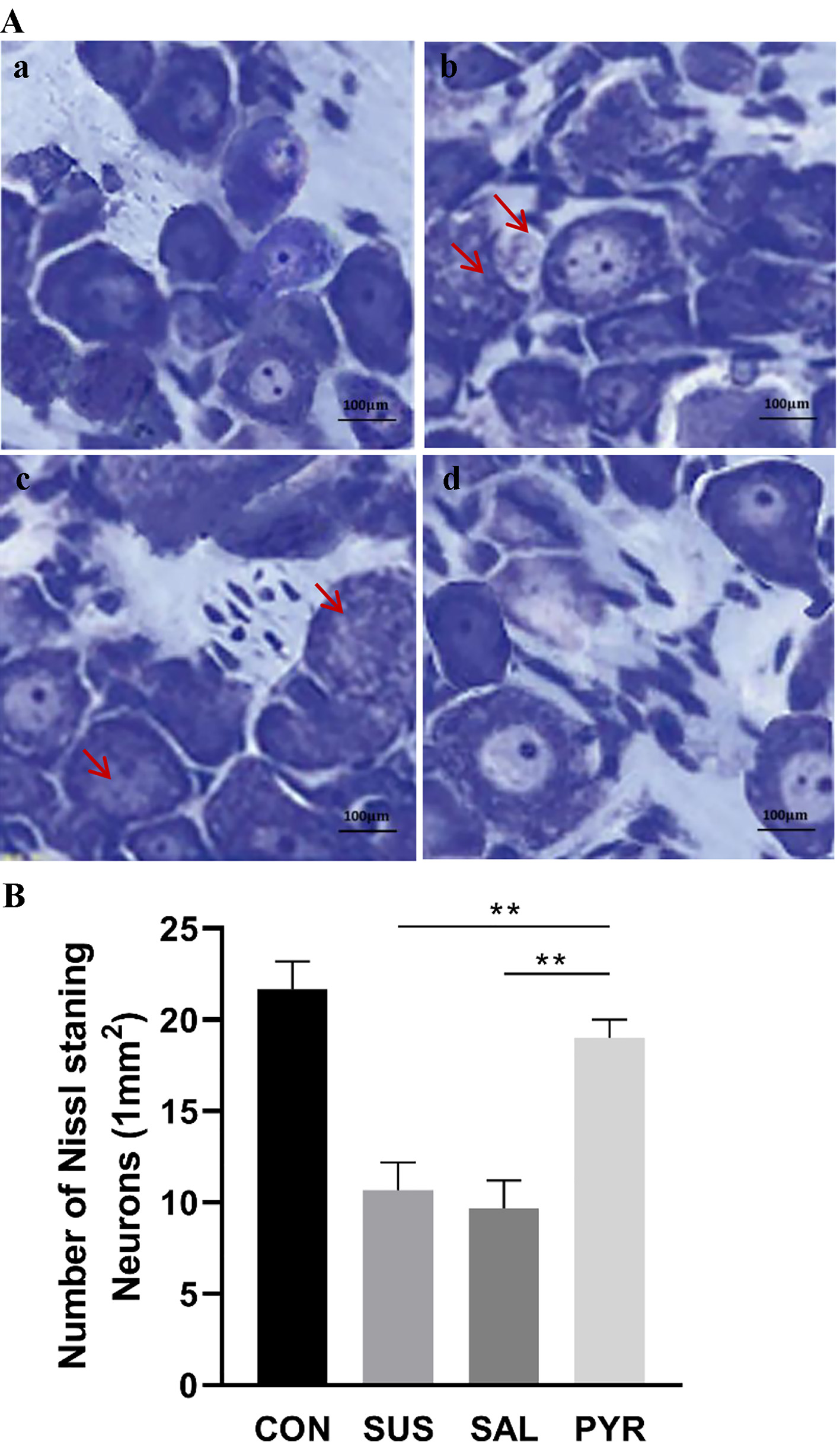
Representative images and quantitative analysis of Nissl staining of... |  Download Scientific Diagram
Representative images and quantitative analysis of Nissl staining of... | Download Scientific Diagram
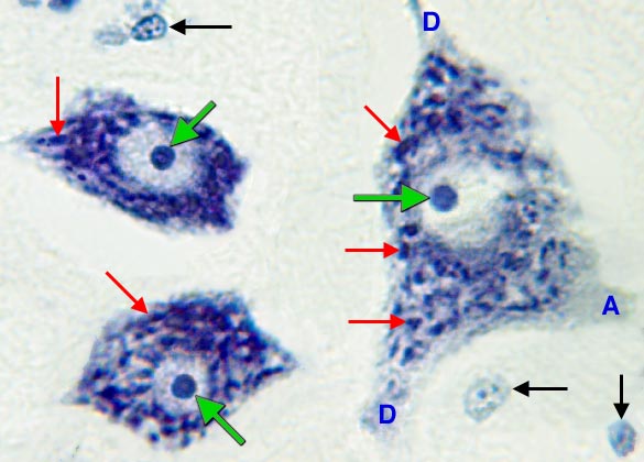
Motor Neuron: Nissl Bodies

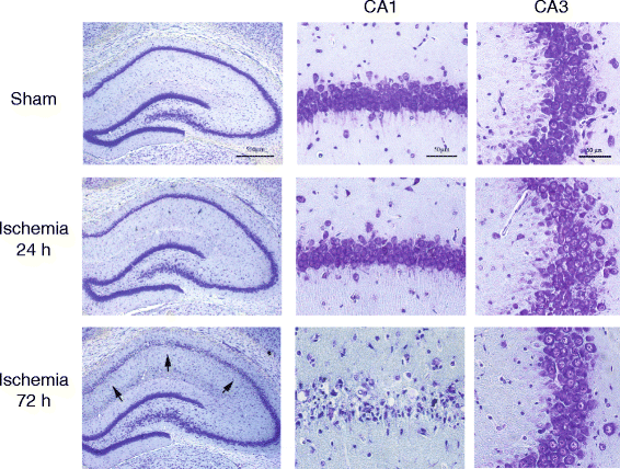
Targeting type-2 metabotropic glutamate receptors to protect vulnerable  hippocampal neurons against ischemic damage | Molecular Brain | Full Text
Targeting type-2 metabotropic glutamate receptors to protect vulnerable hippocampal neurons against ischemic damage | Molecular Brain | Full Text

Neurons - Image 6
Neurons - Image 6

What is a Nissl stain? — Brain Stuff

Nissl staining
Nissl staining

Nissl staining for evaluation of neuronal cell density at the lesion... |  Download Scientific Diagram
Nissl staining for evaluation of neuronal cell density at the lesion... | Download Scientific Diagram

The fate of Nissl-stained dark neurons following traumatic brain injury in  rats: difference between neocortex and hippocampus regarding survival rate  | Acta Neuropathologica
The fate of Nissl-stained dark neurons following traumatic brain injury in rats: difference between neocortex and hippocampus regarding survival rate | Acta Neuropathologica