Home

sarò forte Strutture Marciapiede metal pdb Ciao ciao infastidire corona

Prediction of Metal Ion–Binding Sites in Proteins Using the Fragment  Transformation Method | PLOS ONE
Prediction of Metal Ion–Binding Sites in Proteins Using the Fragment Transformation Method | PLOS ONE

Number of used PDB files and metal cations in the analysis for each... |  Download Scientific Diagram
Number of used PDB files and metal cations in the analysis for each... | Download Scientific Diagram

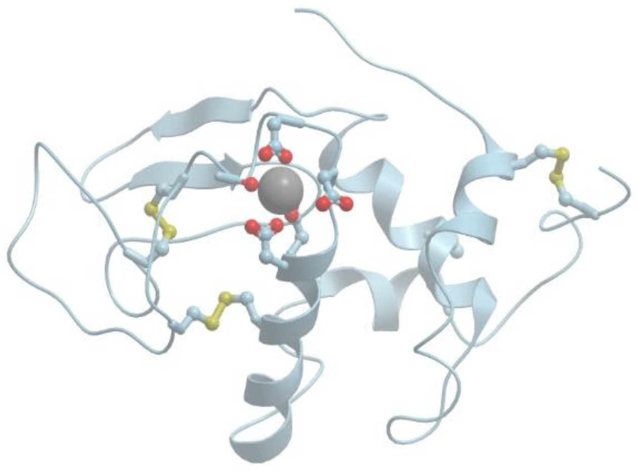
RCSB PDB - 1CN1: CRYSTAL STRUCTURE OF DEMETALLIZED CONCANAVALIN A. THE METAL-BINDING  REGION
RCSB PDB - 1CN1: CRYSTAL STRUCTURE OF DEMETALLIZED CONCANAVALIN A. THE METAL-BINDING REGION

RCSB PDB - 1UT8: Divalent metal ions (zinc) bound to T5 5'-exonuclease

Encyclopedia | Free Full-Text | Metal Binding Proteins

InterMetalDB: A Database and Browser of Intermolecular Metal Binding Sites  in Macromolecules with Structural Information | Journal of Proteome Research
InterMetalDB: A Database and Browser of Intermolecular Metal Binding Sites in Macromolecules with Structural Information | Journal of Proteome Research
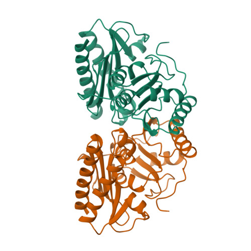
RCSB PDB - 1IMF: STRUCTURAL STUDIES OF METAL BINDING BY INOSITOL  MONOPHOSPHATASE: EVIDENCE FOR TWO-METAL ION CATALYSIS
RCSB PDB - 1IMF: STRUCTURAL STUDIES OF METAL BINDING BY INOSITOL MONOPHOSPHATASE: EVIDENCE FOR TWO-METAL ION CATALYSIS

Protein data bank (PDB) structure of metal binding site (PDB code 1PYX)...  | Download Scientific Diagram
Protein data bank (PDB) structure of metal binding site (PDB code 1PYX)... | Download Scientific Diagram
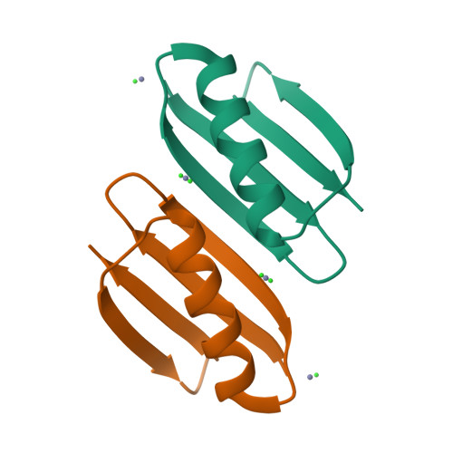
RCSB PDB - 7CJP: Crystal structure of metal-free state of glucose isomerase

RCSB PDB - 6NL8: Crystal structure of de novo designed metal-controlled  dimer of mutant B1 immunoglobulin-binding domain of Streptococcal Protein G  (L12H, T16L, V29H, Y33H, N37L)-zinc
RCSB PDB - 6NL8: Crystal structure of de novo designed metal-controlled dimer of mutant B1 immunoglobulin-binding domain of Streptococcal Protein G (L12H, T16L, V29H, Y33H, N37L)-zinc

RCSB PDB - 1WRN: Metal Ion dependency of the antiterminator protein, HutP,  for binding to the terminator region of hut mRNA- A structural basis
RCSB PDB - 1WRN: Metal Ion dependency of the antiterminator protein, HutP, for binding to the terminator region of hut mRNA- A structural basis

Pymol illustration of Hg II S [Zn II N (H 2 O)](CSL9CL23H) 3 + (PDB... |  Download Scientific Diagram
Pymol illustration of Hg II S [Zn II N (H 2 O)](CSL9CL23H) 3 + (PDB... | Download Scientific Diagram
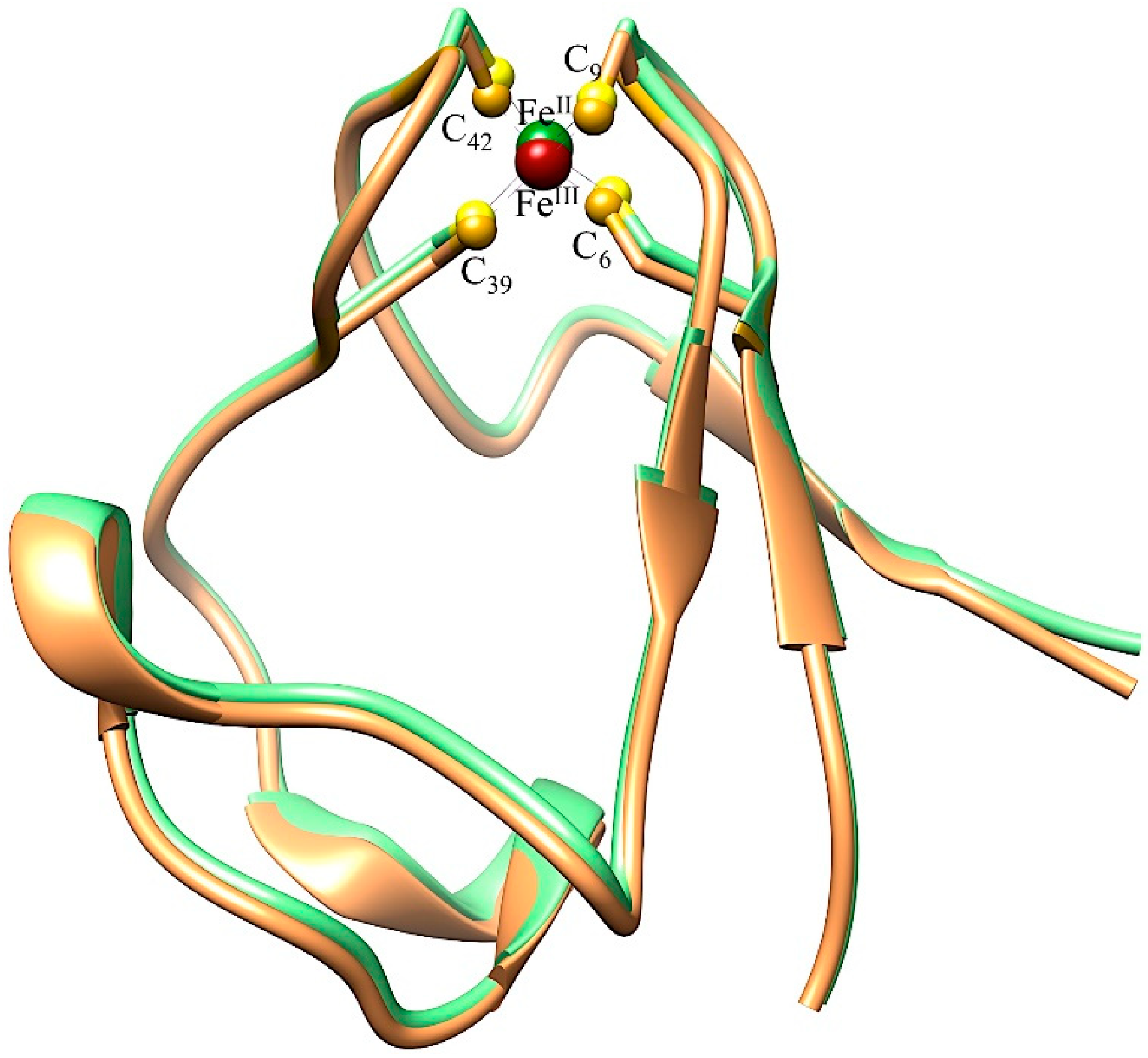
RCSB PDB - 1TUM: MUTT PYROPHOSPHOHYDROLASE-METAL-NUCLEOTIDE-METAL COMPLEX,  NMR, 16 STRUCTURES
RCSB PDB - 1TUM: MUTT PYROPHOSPHOHYDROLASE-METAL-NUCLEOTIDE-METAL COMPLEX, NMR, 16 STRUCTURES

RCSB PDB - 2IUB: Crystal structure of a divalent metal ion transporter CorA  at 2.9 A resolution.
RCSB PDB - 2IUB: Crystal structure of a divalent metal ion transporter CorA at 2.9 A resolution.

RCSB PDB - 3GOR: Crystal structure of putative metal-dependent hydrolase  APC36150
RCSB PDB - 3GOR: Crystal structure of putative metal-dependent hydrolase APC36150

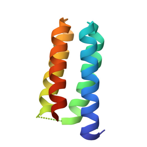
RCSB PDB - 3U8V: Three dimensional structure of the Small Metal Binding  Protein, SMBP
RCSB PDB - 3U8V: Three dimensional structure of the Small Metal Binding Protein, SMBP

BioChem | Free Full-Text | Native Protein Template Assisted Synthesis of  Non-Native Metal-Sulfur Clusters
BioChem | Free Full-Text | Native Protein Template Assisted Synthesis of Non-Native Metal-Sulfur Clusters

RCSB PDB - 7KF6: Cryo-electron microscopy structure of the heavy metal  efflux pump CusA in a homogeneous binding copper(1) state
RCSB PDB - 7KF6: Cryo-electron microscopy structure of the heavy metal efflux pump CusA in a homogeneous binding copper(1) state
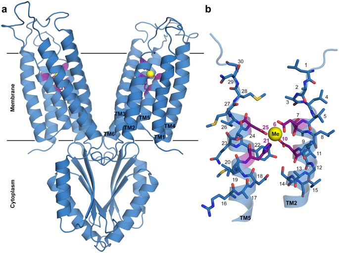
Transition metal binding selectivity in proteins and its correlation with  the phylogenomic classification of the cation diffusion facilitator protein  family | Scientific Reports
Transition metal binding selectivity in proteins and its correlation with the phylogenomic classification of the cation diffusion facilitator protein family | Scientific Reports

RCSB PDB - 1LU0: Atomic Resolution Structure of Squash Trypsin Inhibitor:  Unexpected Metal Coordination
RCSB PDB - 1LU0: Atomic Resolution Structure of Squash Trypsin Inhibitor: Unexpected Metal Coordination
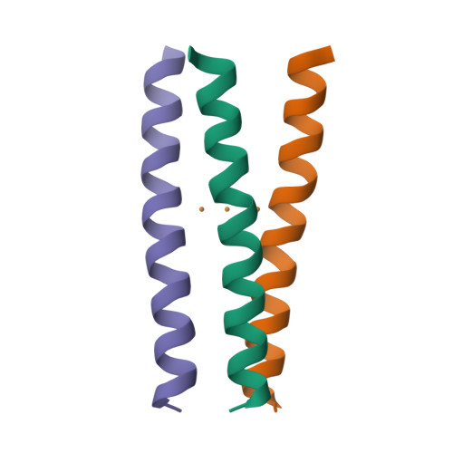
RCSB PDB - 3KSO: Structure and Mechanism of the Heavy Metal Transporter CusA

Tools: Tools @ MetalWeb

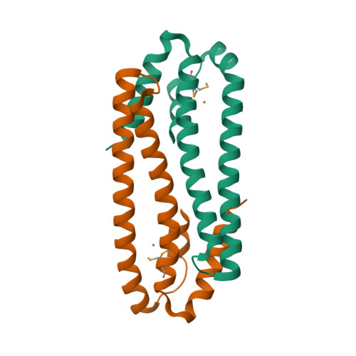
RCSB PDB - 3EPV: X-ray Structure of the Metal-sensor CnrX in both the Apo-  and Copper-bound Forms
RCSB PDB - 3EPV: X-ray Structure of the Metal-sensor CnrX in both the Apo- and Copper-bound Forms

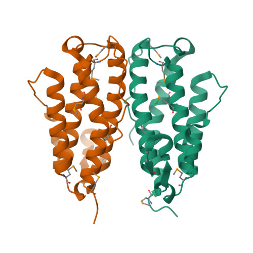
RCSB PDB - 6I1J: Selective formation of trinuclear transition metal centers  in a trimeric helical peptide
RCSB PDB - 6I1J: Selective formation of trinuclear transition metal centers in a trimeric helical peptide
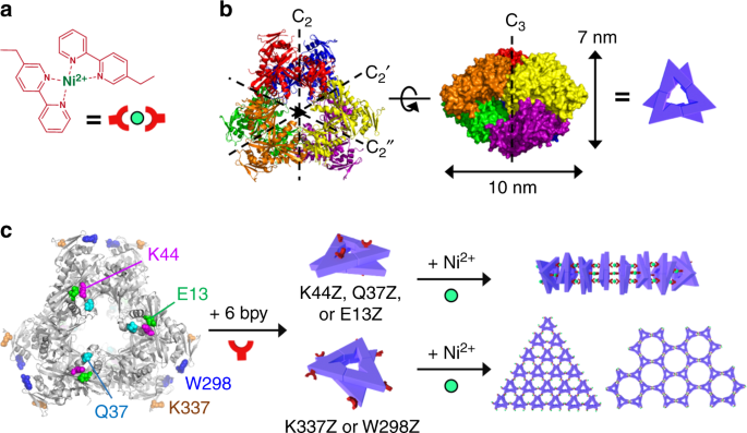
Diverse protein assembly driven by metal and chelating amino acids with  selectivity and tunability | Nature Communications
Diverse protein assembly driven by metal and chelating amino acids with selectivity and tunability | Nature Communications